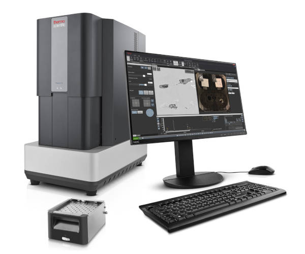
Phenom ParticleX Steel Desktop SEM
Desktop SEM enabling high quality steel manufacturing through failure analysis and process improvement
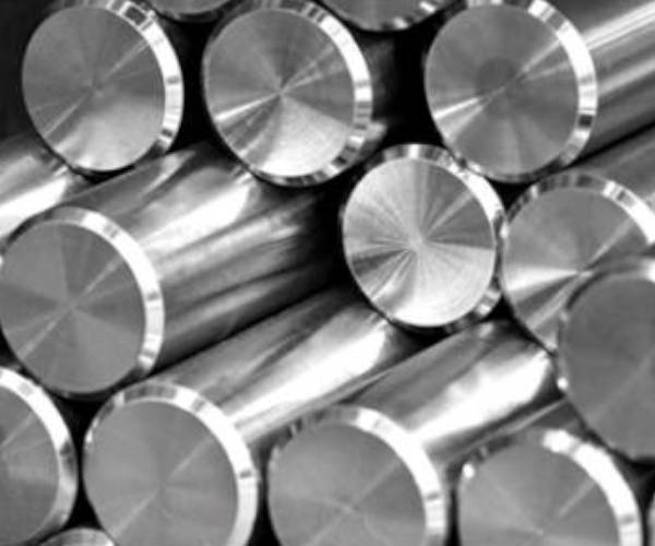
Steel metallurgists and researchers rely on scanning electron microscopy (SEM) and energy dispersive X-ray spectroscopy (EDS) for critical tasks like failure analysis and process optimization. The Thermo Scientific Phenom ParticleX Steel Desktop SEM provides a versatile solution, enabling both detailed failure analysis and automated characterization of non-metallic inclusions within steel samples.
This versatile solution delivers high-quality imaging and elemental analysis for steel samples, supporting efficient production of high-value steels. Its rapid analysis capabilities enable swift responses to customer claims, while automated inclusion analysis provides valuable insights into the steelmaking process.
This versatile solution delivers high-quality imaging and elemental analysis for steel samples, supporting efficient production of high-value steels. Its rapid analysis capabilities enable swift responses to customer claims, while automated inclusion analysis provides valuable insights into the steelmaking process.
REQUEST A QUOTE / ENQUIRY FORM
Features
(ParticleX Steel Desktop SEM – Workflow introduction)
(Phenom ParticleX Steel analysis of inclusions)
Small Footprint
The Phenom ParticleX Steel Desktop SEM uses standard wall power, eliminating the need for infrastructure changes. Its integrated EDS provides ‘click-and-go’ elemental mapping and line scans for quantified element distribution.
Ease of use
Building on the intuitive interface of the established Phenom Desktop SEMs, this system ensures a minimal learning curve for both experienced and new users. The high-brightness CeB6 source enhances image detail and accelerates automated steel inclusion analysis, maximizing efficiency.
Future-proof
Although the system comes with pre-set classification rules and analysis recipes for immediate steel inclusion analysis, it also provides complete customization options. This flexibility allows users to refine their analysis and uncover new insights through updated recipes.
Specifications
| Electron optical magnification range |
|
| Light optical magnification |
|
| Resolution |
|
| Image resolution options |
|
| Acceleration voltages |
|
| Vacuum levels |
|
| Detector |
|
| Sample size |
|
| Sample loading time |
|
PHENOM PARTICLEX DESKTOP SEM
PHENOM PARTICLEX DESKTOP SEM
Menu