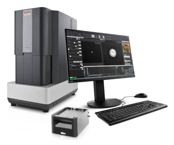
Phenom ParticleX AM Desktop SEM
Desktop SEM for additive manufacturing analysis, capable of observing large samples up to 100 mm x 100 mm.
The Thermo Scientific Phenom ParticleX Desktop Scanning Electron Microscope (SEM) is a versatile SEM specifically designed for additive manufacturing, providing high purity at the microscale.
Its chamber accommodates samples as large as 100 mm x 100 mm. The innovative venting and loading mechanism ensures the quickest vent/load cycle in the industry, maximizing throughput.
Its chamber accommodates samples as large as 100 mm x 100 mm. The innovative venting and loading mechanism ensures the quickest vent/load cycle in the industry, maximizing throughput.
With the Phenom ParticleX AM Desktop SEM, you gain full control of your data in-house:
- Monitor essential characteristics of metal powders
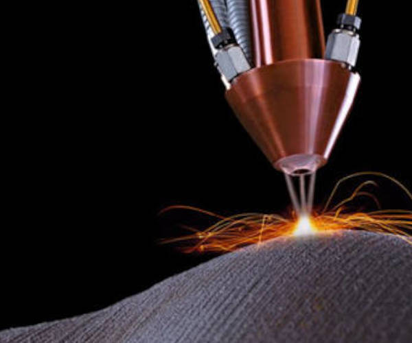
- Optimize powder-bed and powder-fed additive manufacturing processes
- Identify particle size distributions, individual particle morphology, and foreign particles.
REQUEST A QUOTE / ENQUIRY FORM
Features
Additive manufacturing testing
The Phenom ParticleX AM Desktop SEM measures particle size and shape (diameter, perimeter, aspect ratio, roughness, Feret diameter) and displays results as d10, d50, and d90 values.
SEM particle analysise
The Phenom ParticleX AM Desktop SEM offers a 100 mm x 100 mm chamber with a fast motorized stage. Its proprietary loading shuttle delivers a 60-second vent/load cycle, providing superior throughput.
SEM elemental mapping
Single-click elemental mapping and line scans provide quantified element distribution via a line plot, useful for analyzing edges, coatings, and multilayer samples.
Secondary electron detector
The optional SED on the Phenom ParticleX AM Desktop SEM provides detailed surface information by collecting low-energy electrons, useful for topography and morphology analysis, especially for microstructures, fibers, and particles.
Specifications
| Electron optical |
|
| Electron optical magnification range |
|
| Light optical magnification |
|
| Resolution |
|
| Image resolution options |
|
| Acceleration voltages |
|
| Vacuum levels |
|
| Detector |
|
| Sample size |
|
| Sample loading time |
|
PHENOM PARTICLEX DESKTOP SEM
Menu