Leica THUNDER Imager Tissue
Decode 3D biology in real time *
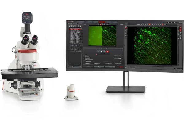
The THUNDER Imager Tissue allows real-time fluorescence imaging of 3D tissue sections typically used in neuroscience and histology research. Acquire rich, detailed images of thick tissues free of haze from out-of-focus blur.
Even fine structures deep in tissues can be resolved thanks to Computational Clearing, an innovative Leica technology. Image detailed morphological structures like axons and dendrites of neurons in a brain slice. The high image quality, even with thick tissue sections, is combined with the well-known speed, fluorescence efficiency, and ease of use of widefield microscopes.
Gain these advantages using a THUNDER Imager Tissue for your research:
- Get computationally cleared images directly in your live preview with THUNDER Live
- Rapidly acquire blur-free images showing finest details of the morphology, even deep within thick sections
- Get fast overviews of whole tissue sections
- Image and analyze challenging tissue sections with an easy workflow
REQUEST A QUOTE / ENQUIRY FORM
Begin every experiment with confidences
Perform high resolution tissue imaging on thick samples without struggling to find your region of interest.
The new THUNDER Live add-on visualizes a computationally cleared image instantly in the live viewer and allows you to optimize ICC parameters by using live image feedback.
Your benefits with THUNDER Live:
The new THUNDER Live add-on visualizes a computationally cleared image instantly in the live viewer and allows you to optimize ICC parameters by using live image feedback.
Your benefits with THUNDER Live:
- Get computationally cleared images directly in your live preview.
- Reduce the time to optimal results with immediate visual feedback.
- Intuitively and quickly find the best THUNDER ICC parameters on the fly while still in live view.
- Optimize your image in one simple step without changing any settings.
- THUNDER Live’s blur-free view also makes selecting significant regions of interest in your sample a breeze, even on thick specimens.
Speed up tissue imaging
Increase your slide scanning experiment speed and perform fast and efficient multicolor acquisiton. You’ll be able to assess more markers with less photobleaching.
Your advantages with the accelerated tissue imaging
Your advantages with the accelerated tissue imaging
- Scan your slides faster with a synchronized experiment control
- Multicolor acquisitions are more than two times faster when using the Synapse controller
- Extract contextual information unbelievably fast in slide scanning multicolor experiments
- Evaluate more markers through precise excitation control of multiline LED light sources with less photobleaching
Resolve fine details in challenging specimens
THUNDER Imager Tissue delivers instantly cleared-up fluorescent images with fine details of your multi-color tissue sections. Just turn the THUNDER Imager on and get started! The unique Computational Clearing uses optimal parameters to produce expert-level results. It does it automatically with no need for calibration or user interaction.
Achieve optimal fluorescence and contrast settings instantly via the patented Leica fluorescence intensity (FIM) and contrast managers. Choose from a range of objectives, optimized for specific applications, to ensure outstanding results even for challenging specimens.
Achieve optimal fluorescence and contrast settings instantly via the patented Leica fluorescence intensity (FIM) and contrast managers. Choose from a range of objectives, optimized for specific applications, to ensure outstanding results even for challenging specimens.
THUNDER Imager 3D Tissue
Two Options for THUNDER Imager Tissue
Choose the configuration which best meets your requirements:
- THUNDER Imager 3D Tissue is a fully automated tissue imaging system for recording multi-color 3D images, now directly in your live preview*. Capture multiple images in the z-direction and visualize them in the 3D viewer. Achieve excellent z-stack imaging due to a precise motorized focus drive.
- For brilliant imaging of single planes and fast overviews of your tissue the THUNDER Imager Tissue is also available in a cost-effective configuration without a motorized focus drive.
Combination System: LMD System with THUNDER Imaging
THUNDER can be combined with additional Laser Microdissection (LMD) capabilities in the same system. The base stand of the LMD systems and THUNDER Imager 3D Tissue are the same, thus such a combination offers:
- Lab space savings with one system for different tasks
- Brilliant fluorescence imaging with THUNDER using LAS Laser Microdissection with the
- LMD option using the unique LMD Software to visualize and mark regions of interest for subsequent dissection and collection via gravity into standard vials ready for downstream processing such as RNAseq, NGS, MS, qPCR, microarray etc.
TECHNICAL SPECIFICATIONS
| THUNDER Imager TissueBrilliant imaging of single planes and fast overviews | THUNDER Imager 3D TissueFully automated tissue imaging system for recording multi-color 3D images | |
| Microscope Platform | DM4 B | DM6 B |
| Display/Touch screen | Display shows all microscope settings at a glance | SmartTouch controls the microscope conveniently and intuitively |
| Focus | Mechanical | Motorized |
| Stage | Motorized with integrated controller, mechanical (optional) | Motorized or scanning stages |
| LAS | Yes (optional) | Yes |
| Transmitted Light Illumination | LED | LED |
| Transmitted Light Contrast Methods | BF, optional PH, DF, POL, DIC | BF, optional PH, DF, POL, DIC |
| Fluorescence Axis | Fully automated | Fully automated, with Internal Filter Wheel (IFW) and Excitation Manager |
| Fluorescence Illumination | EL6000 (metal halide), LED | LED, EL6000 (optional) |
UPRIGHT LIGHT MICROSCOPE
Menu