Leica Visoria M Materials Microscope
Achieve greater efficiency and comfort in your daily microscopy tasks. The Visoria B laboratory microscope is designed specifically for life science and clinical laboratory applications.
Simplify your workflow with encoded functions, optimized lighting, and a range of advanced features. Its ergonomic design also helps you work more comfortably, reducing strain during extended use.
REQUEST A QUOTE / ENQUIRY FORM
(Visoria M materials microscope)
Introducing Visoria M
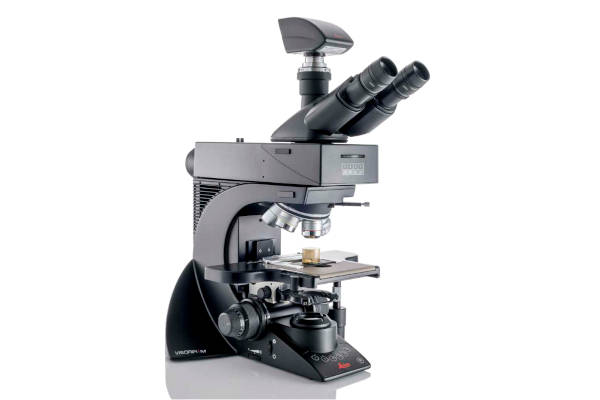
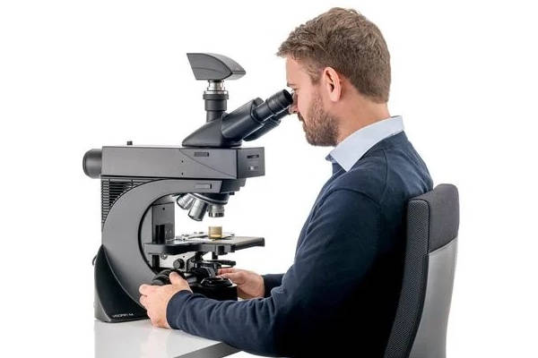
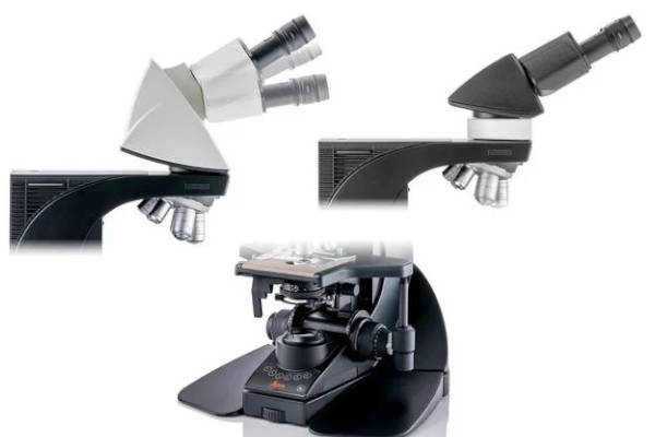
Your solution for routine material inspection
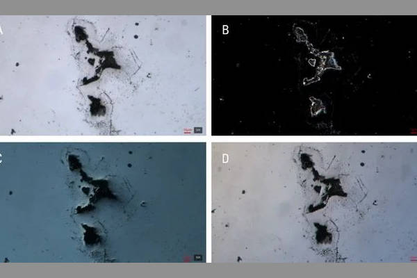
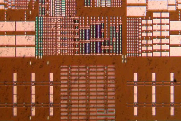
The Visoria M materials microscope enables detailed examination of the microstructure of metals, alloys, electronic and mechanical components, composite materials, glass, ceramics, and more. When paired with the Enersight software platform, it also allows for precise cross-section and layer-thickness analysis.
(Observe samples with optimal illumination without the need to adjust the microscope’s light settings)
Save time with optimized lighting settings
With Visoria M, you can focus more on observing and analyzing your samples. When you adjust the microscope’s magnification or contrast mode, there’s no need to manually tweak the brightness. Thanks to the light management feature and built-in encoding, the illumination settings are automatically adjusted for you.
(Save time and effort by snapping your image with a press of a button on the microscope stand.)
Streamline Your Documentation
Effortlessly capture sample details with a simple press of a button, all while keeping your focus on the image. The image acquisition button is conveniently located on the Visoria M microscope stand for easy access.
When you save an image, the system automatically records selected settings along with the image’s metadata.
Additionally, the scale bar is automatically adjusted and added to the image, boosting efficiency and saving you valuable time.
When you save an image, the system automatically records selected settings along with the image’s metadata.
Additionally, the scale bar is automatically adjusted and added to the image, boosting efficiency and saving you valuable time.
(The aperture diaphragm’s scale on the condenser has color markings matching the objective color codes.)
Effortless Microscope Operation
Carry out your daily tasks quickly and confidently with the user-friendly design of the Visoria M.
- Easily identify the correct aperture for each objective using the convenient color-coding system.
- Safeguard your samples and objectives from accidental damage with the integrated focus stop.
- Achieve precise focusing at higher magnifications with the three-gear focusing system, offering coarse, medium, and fine adjustments
(User operating the Visoria M digital material microscope)
Work without eyepieces by going digital
The Visoria M digital material microscope without eyepieces offers a number of practical benefits:
- Enjoy a comfortable and relaxed working posture by viewing images directly on a tablet.
- Quickly visualize and document your workflow, making it easy to share and discuss image results with colleagues.
- Save valuable workspace since no computer is required.
(Users can maintain a comfortable position while working with Visoria M)
Maintain Comfort Throughout Your Workday
Visoria M is designed to support your well-being, promoting proper posture and helping to minimize neck and back strain during extended microscope sessions.
Enjoy comfortable operation with ergonomically aligned shoulders and hands, thanks to the symmetrical design and adjustable height of the focus and stage control knobs. You can easily operate the Visoria M with just one hand.
Switch effortlessly between right- and left-handed use, making it ideal for shared workspaces and multiple users.
(Flexibly adapt the eyepiece viewing angle and height as well as the overall microscope height)
Customize your microscope with ergonomic accessories
Visoria B’s adaptable design helps you maintain an upright posture. Select from a variety of ergonomic accessories to match your preferences:
- Ergonomic tubes: Opt for 15° ergonomic tubes or adjustable VarioTubes (0–35° tilt) for a relaxed head position and flexible viewing angles.
- Ergonomic modules: Add ErgoModules beneath the tube to set the eyepiece height for comfortable seated use.
- Ergonomic lift: The optional ErgoLift makes it easy to adjust the microscope’s height for optimal comfort.
(The XY stage position and focus knobs are easily accessible and adjustable with one hand)
Minimize strain with less repetitive movement
If you spend long hours at the microscope, Visoria B helps reduce discomfort and the risk of repetitive strain injuries. Adjust the height and torque of the stage and focus controls to minimize repetitive motions and enhance your comfort during extended use.
(Images of the same inclusions acquired with A) brightfield, B) darkfield, C) DIC, and D) oblique illumination using Visoria M)
See Every Detail with Optimal Contrast
For industrial inspections, materials analysis, and R&D, Visoria M enables you to clearly observe structural features and detect defects like scratches or contamination on your samples.
The microscope offers a range of contrast techniques, including:
- Brightfield
- Darkfield
- Polarization
- Differential Interference Contrast (DIC)
- Oblique Illumination
- Fluorescence
(Save time when screening samples using the optional 0.7x Macro objective for a fast overview)
Get a Rapid Sample Overview with the 0.7x Macro Objective
When you need to examine larger structures, the optional 0.7x Macro objective lets you quickly transition from a broad sample overview to detailed observation. With a field of view of approximately 36 mm in diameter, you can easily orient yourself and gain a comprehensive overview in less time compared to conventional objectives—making sample screening more efficient.
For closer inspection, simply switch to higher magnification using the wide selection of available objectives to reveal fine sample details.
(The semi-automated measurement provides the minimum, maximum, and average thickness information of a coating or layer)
Enhanced by the Enersight Software Platform
Make your workflow smoother and more efficient with the combination of the Visoria M materials microscope and the Enersight software platform. This intuitive interface allows you to easily compare, measure, and share your data all in one place.
Key Benefits:
- Accurately determine coating or layer thickness with the Layer Thickness Measurement feature.
- View samples in greater detail with a wider field of view and higher resolution using the XY Stitching with Manual Stage function.
- Capture crisp, clear images with extended depth of field (EDOF).
- Achieve optimal image quality by utilizing the Quick Brightness function for perfect illumination and camera settings.
- Automatically correct shading caused by uneven illumination for consistently high-quality images.
- Deepen your understanding of samples by merging images taken with different contrast methods, such as brightfield and darkfield.
UPRIGHT LIGHT MICROSCOPE
Menu

